 |
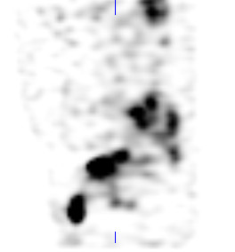
| Fig. 1 Total body bone scan of a patient diagnosed with renal cell carcinoma. |
As a technologist what would you do after the total body bone scan (TBBS) was complete? I think one of our main duties are to review the images to see if there are any asymmetrical bone uptake relating to the patient's history and to take extra pictures if needed. With respect to the images above, there are a few spots, in particular the spine, but other than that nothing too remarkable to talk about. Unless you take a closer look at the pelvis. Is this contamination or is this "something else"? Well it has to be something else or else it wouldn't be on this blog.
So, what are our options to figure out, what's going on in the pelvis?
a. Take a quick lateral view, to see if this is truly contamination
b. SPECT or SPECT/CT
c. Squat view on the camera (ie. have the patient sit on the camera) to separate bone versus soft tissue
Since we have a hybrid system, we SPECT/CT'ed the pelvis. So the question you are asking yourself right now is, why did we opt to do this? Well if you look at urine contamination on patients in general, it usually looks more "drawn out" - dribbling with smaller spots. Also in this TBBS the margins of the "spot" below the bladder proper, are smooth and well defined... so it doesn't really fit with our pre notion of what contamination would look like. Plus, we had the time and we needed the "billings" and the workload units, so we did it... I am joking of course (as someone pointed out) .... And this is what we got:
The SPECT is interesting. The "spot" is anterior to the bladder, but really not on the surface of the skin. Below is the CT transaxial slice of the pelvis from the SPECT/CT.
Notice the bladder in the pelvis (click on link for normal looking bladder). It looks like a "pear" with the most anterior lobe shifted to the right extending beyond the pubic bone. Is this still contamination? Here's another look with the sagittal slices for SPECT, CT and fused SPECT/CT slices.
Well if you haven't figured it out now, the "spot" that you actually see is the bladder. The bladder has herniated out and dropped below the pelvic floor... the patient has a herniated bladder! This is something that we normally do not see very often.
The indication for this patient was to have a metastatic work up, because they were re-diagnosed with renal cell carcinoma (RCC), this time in the right kidney. The bone scan was included in the work up for metastases in the bones. There were no clear cut evidence for this, but there were a lot of expressions of degeneration in the joints and notice one kidney is missing... the left one on the TBBS. The left kidney had been removed in 2009 as the result of RCC. Even though there were no bony evidence of metastases, the separate CT scans showed a large liver lesion indicating the progression of his disease from his right kidney (images not shown).
So, what are our options to figure out, what's going on in the pelvis?
a. Take a quick lateral view, to see if this is truly contamination
b. SPECT or SPECT/CT
c. Squat view on the camera (ie. have the patient sit on the camera) to separate bone versus soft tissue
Since we have a hybrid system, we SPECT/CT'ed the pelvis. So the question you are asking yourself right now is, why did we opt to do this? Well if you look at urine contamination on patients in general, it usually looks more "drawn out" - dribbling with smaller spots. Also in this TBBS the margins of the "spot" below the bladder proper, are smooth and well defined... so it doesn't really fit with our pre notion of what contamination would look like. Plus, we had the time and we needed the "billings" and the workload units, so we did it... I am joking of course (as someone pointed out) .... And this is what we got:
The SPECT is interesting. The "spot" is anterior to the bladder, but really not on the surface of the skin. Below is the CT transaxial slice of the pelvis from the SPECT/CT.
| Fig. 2 Transaxial CT image of the pelvis from the SPECT/CT scan. Notice the "pear" shaped bladder. |
Notice the bladder in the pelvis (click on link for normal looking bladder). It looks like a "pear" with the most anterior lobe shifted to the right extending beyond the pubic bone. Is this still contamination? Here's another look with the sagittal slices for SPECT, CT and fused SPECT/CT slices.
 |
| Fig. 3 Sagittal SPECT view of the pelvis. Notice the bladder and the anterior ovoid "spot". |
 |
| Fig. 4 Sagittal CT slice of the pelvis. The bladder has a flaccid phallic appearance. |
 |
| Fig. 5 Fused SPECT/CT sagittal slice. Everything is starting to make a little bit more sense. |
Well if you haven't figured it out now, the "spot" that you actually see is the bladder. The bladder has herniated out and dropped below the pelvic floor... the patient has a herniated bladder! This is something that we normally do not see very often.
The indication for this patient was to have a metastatic work up, because they were re-diagnosed with renal cell carcinoma (RCC), this time in the right kidney. The bone scan was included in the work up for metastases in the bones. There were no clear cut evidence for this, but there were a lot of expressions of degeneration in the joints and notice one kidney is missing... the left one on the TBBS. The left kidney had been removed in 2009 as the result of RCC. Even though there were no bony evidence of metastases, the separate CT scans showed a large liver lesion indicating the progression of his disease from his right kidney (images not shown).
Aside from the reasons given, I was surprised that you also mentioned exposing patients to CT because you needed the "billing" and workload units.
ReplyDeleteI am kidding of course... our doctors wanted to ensure that this was indeed bladder and not something else. The previous CT did not indicate any herniation.
DeleteAlthough newest technologies provide greater information...back in 1982 "The Giga Squat View" was used to delineate pelvic radionuclide uptake. Ref: Journal of Nuclear Medicine Technology, Vol 10, No.1 March '82
Delete