 |
| Fig. 1 This is an anterior TBBS image, but something doesn't seem quite right. |
 |
| Fig.2 The posterior TBBS image. Expand the image. |
When we first looked at these images we were unsure what the problem was, but the images did not look right. The total body bones scan was taken on a Philips ADAC Skylight system, but generally the images do not look this way. This is what I mean.... the images look hazy and not indicative of a normal quality bone scan. Some of you may have figured it out by now, but for those who are still guessing, it was caused by a student.
The patient had come to the Nuclear Medicine department to determine if osteomyelitis had developed in the right shoulder. They have had several corrective surgeries to their joints already: bilateral shoulder replacement, bilateral hip replacement and a left knee replacement. Let's put it this way, when she came into the department the patient was held together by duct tape... joking of course but the patient was in pain.
However knowing her condition, we had to repeat the images because something was amiss. Check out the images below, which were also performed on the Philips ADAC system.
 |
| Fig. 3 Corrected TBBS images. Compare to Fig. 1 and Fig. 2. Expand the image. |
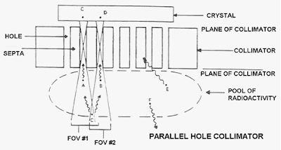
So the problem is this... when the student had placed the patient on the bed, they had unknowingly changed the collimators to high energy collimators instead of the high resolution collimators (VXGP). While setting up the patient in the computer for the acquisition, the computer had prompted the student that the wrong collimators were installed. Majority of our acquisition protocols are customized to the organ system that we are scanning (like most modern departments), with a predetermined energy spectrum and energy window, collimation, length of time for the scan etc.. In this case the student did not bother to read the prompt which notified that the wrong collimators were installed for this particular acquisition protocol. They ignored it and proceeded with the scan. Thus the 140 keV gamma rays hitting the crystal were limited by the thicker collimation of the high energy collimators, since they were designed for high energy isotopes to minimize the amount of cross talk. Collimation is one of the fundamental principles of Nuclear Medicine Instrumentation.
Another possible way of getting images to look like those in Fig. 1 and Fig. 2 is by having the wrong energy or energy window settings. Although a lot of the camera systems all now have presets, but it's always a good habit to check your parameters and never ignore your error messages.
In the end this patient had their total body bone scan, along with their In-111 WBC with sulphur colloid two days later and found no osteomyelitis in the right shoulder.

No comments:
Post a Comment