Showing posts with label Bone imaging. Show all posts

Thursday, 6 February 2014

Erdheim Chester Disease

A lot of times when you read a requisition, you come across a lot of diseases, syndromes and terms, especially acronyms, that medical personnel use to which you have no clue what they mean sometimes.  If it piques your interest you would look them up and if it is really pertinent to the case you would pay attention to what is written.

Well reading Erdheim Chester disease on the requisition, I really did not give a second thought (just another disease process), until I saw this.


Fig. 1  Total body bone scan of a patient with Erdheim Chester disease.  The patient had complained of localized pain in the femurs.
Fig. 2  Additional images of the arms, for completeness. 
Fig. 3 Additional images of arms to document both dorsal and ventral views... I know, I know... I didn't label them, and the fingers were cut off on the view, but they didn't really involve in completing the whole case.
So what is Erdheim Chester Disease (ECD)?  Click on the link and find out a little more, but what is interesting is that pathologists do not know how to classify this disease because it is so rare.  The bottom line is, it is a disease in which there is an accumulation of histiocytes depositing themselves into loose connective tissue.  It eventually causes thickening and progresses into dense fibrotic tissue over time.  The symptoms tend to be variable, but the one presented in this case was discomfort in the thighs and femurs, fatigue and intermittent back pain. We performed a flow and blood pool sequence of the lower body and there was a marked increase in perfusion in the blood flow and blood pool in the distal femurs*.


Fig. 3  Blood pool image after the injection of Tc99m-MDP.  The distal femurs demonstrate an increased perfusion.
The patient overall seemed healthy, when interviewed during the examination, except for the minor complaints mentioned above.  The images were checked with the radiologist upon completion of the bone scan and was released.  

So at the end of the day the lesson learned is, not to dismiss diseases, syndromes or terms so readily because it may provide an opportunity to learn.

*Note:  When we performed the total body bone scan we saw what was presented and then went back to review the flow and blood pool study more closely. 

Thursday, 17 May 2012

Bone Scans and CPR

Fig. 1 Patient presents with multiple rib fractures as well as a fracture of the sternum

So what's wrong with this picture?  There are several things as you can note with multiple rib fractures and a sternal fracture as well.  Originally the patient was to have scans on the hands and wrist as indicated on the requisition.  A total body  scan was performed with locals of the hands and wrist.  The locals were unremarkable, but the chest and ribs are very remarkable.

Some clues for you.  No recent thoracic surgery or recent motor vehicle accident.  The patient walked into the department with no deficits.  However, and this might give it away, he did recently have a heart attack.

If you have guessed it, this person was a recipient of CPR administration a month earlier after he had a heart attack.  The delivery of chest compressions from the CPR contributed to the current presentation.  If this was me, forget the CPR, just use the AED's and zap away!

Click here for another example that I found online in regards to bone scans and CPR.

Thursday, 3 May 2012

McCune Albright Syndrome


Sorry about the appearance of the image... I was not able to invert the colour scheme.

However, this patient presented to our department to determine if osteomyelitis was present in her right upper jaw.  In short, our doctors were unable to decide if the site had osteomyelitis, because of other confounding circumstances such as the patient's recent dental work in the same area.  It was tough because they were unsure if there was some type of odontogenic infection occurring as well.  Plus the extent of the underlying bone that was involved in this infection was difficult to resolve given the patient's underlying bony lesions.  The sulphur colloid (bone marrow images) and the delayed In-111 WBC images are not included for viewing because the interesting point about this case, is the actual total body bone scan itself.

McCune Albright Syndrome (polyostotic fibrous dysplasia) is a genetic disorder that affects the bones and pigmentation of the skin.  It is not a common disorder and the exact number of cases in the United States and internationally is not known.  However what is interesting are the multiple abnormal foci of increased activity noted within the bones, this in keeping with the nature of the syndrome. Anyway I thought this was more interesting than the osteomyelitis component of this case.

Wednesday, 2 May 2012

A Weird Incidental Finding

Fig. 1  Total body bone scan of a patient diagnosed with renal cell carcinoma.
As a technologist what would you do after the total body bone scan (TBBS) was complete?  I think one of our main duties are to review the images to see if there are any asymmetrical bone uptake relating to the patient's history and to take extra pictures if needed.  With respect to the images above, there are a few spots, in particular the spine, but other than that nothing too remarkable to talk about.  Unless you take a closer look at the pelvis.  Is this contamination or is this "something else"?  Well it has to be something else or else it wouldn't be on this blog.

So, what are our options to figure out, what's going on in the pelvis?
a.  Take a quick lateral view, to see if this is truly contamination
b.  SPECT or SPECT/CT
c.  Squat view on the camera (ie. have the patient sit on the camera) to separate bone versus soft tissue

Since we have a hybrid system, we SPECT/CT'ed the pelvis.  So the question you are asking yourself right now is, why did we opt to do this?  Well if you look at urine contamination on patients in general, it usually looks more "drawn out" - dribbling with smaller spots.  Also in this TBBS the margins of the "spot" below the bladder proper, are smooth and well defined... so it doesn't really fit with our pre notion of what contamination would look like.  Plus, we had the time and we needed the "billings" and the workload units, so we did it... I am joking of course (as someone pointed out) .... And this is what  we got:


The SPECT is interesting.  The "spot" is anterior to the bladder, but really not on the surface of the skin.  Below is the CT transaxial slice of the pelvis from the SPECT/CT.

Fig. 2 Transaxial CT image of the pelvis from the SPECT/CT scan.  Notice the "pear" shaped bladder.

Notice the bladder in the pelvis (click on link for normal looking bladder).  It looks like a "pear" with the most anterior lobe shifted to the right extending beyond the pubic bone.  Is this still contamination?  Here's another look with the sagittal slices for SPECT, CT and fused SPECT/CT slices.
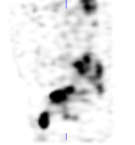
Fig. 3  Sagittal SPECT view of the pelvis.  Notice the bladder and the anterior ovoid "spot".

Fig.  4  Sagittal CT slice of the pelvis.  The bladder has a flaccid phallic appearance.

Fig.  5  Fused SPECT/CT sagittal slice.  Everything is starting to make a little bit more sense.

Well if you haven't figured it out now, the "spot" that you actually see is the bladder.  The bladder has herniated out and dropped below the pelvic floor... the patient has a herniated bladder!  This is something that we normally do not see very often.

The indication for this patient was to have a metastatic work up, because they were re-diagnosed with renal cell carcinoma (RCC), this time in the right kidney.  The bone scan was included in the work up for metastases in the bones.  There were no clear cut evidence for this, but there were a lot of expressions of degeneration in the joints and notice one kidney is missing... the left one on the TBBS.  The left kidney had been removed in 2009 as the result of RCC.  Even though there were no bony evidence of metastases, the separate CT scans showed a large liver lesion indicating the progression of his disease from his right kidney (images not shown).

Wednesday, 21 March 2012

Arterial Injection


What's going on with these total body bone images?  If the title didn't give it away, then you probably know that the Tc99m-MDP injection went into the arterial system rather than into the venous side.  We call it the Michael Jackson sign, for the "gloved one".  A student had injected in the left antecubital fossa ... did they notice that the vein was pulsating rhythmically?  ... not sure.

This is typical of an arterial injection with a high amount of tracer deposition distal to the injection site.  We know that the MDP had been prepared correctly since there are no choroid plexus, thyroid, salivary or gastric uptake that would result from free pertechnetate.  Furthermore bone uptake is visible.  Now as with an interstitial injection with such a large dose, we would normally see uptake in the lymph nodes on the side that the injection was given... but the image did not pick up any lymph nodes (trust me .. I know it's hard to see)

What other conditions may have caused this type of uptake?  Soft tissue uptake in bone scans can result from:


Muscle Uptake on MDP Bone Scan


This is an interesting scan, because not often do we come across muscle uptake in our bone scans.  For the most part we shouldn't see muscle uptake at all, however it is not uncommon, and there has been several journaled articles relating to this fact.  Having "googled" the topic, it appears that there are several conditions that can cause this extra osseous uptake.  There has also even been one case in which exercise prior to the bone scan has caused muscle uptake.

On a quick note, here are some interesting articles (Radionuclide Bone Imaging:  An Illustrative Review) in regards to screening pathological conditions and as well Nonosseous, Nonurologic Uptake on Bone Scintigraphy:  Atlas and Analysis that provides some of the pathways in which Tc99m MDP are involved with non osseous uptake.

With this patient here, you can see if you expand the image, that there are diffuse muscle and soft tissue uptake throughout the body:  hip flexors, quadriceps, calf muscles, deltoids, triceps  and breast tissue.  The patient had been suffering from left sided flank pain and had undergone several examinations to determine the nature of the pain.  This person also has had a previous liver transplant and heart transplant as the result of being diagnosed with glycogen storage disease (GSD) type IIIA at an early age.  We are not really sure how the Tc99m MDP got into the muscles and other soft tissues, but conditions such as polymyositis, myositis ossificans or amyloidosis can cause this appearance.  However there are a slew of conditions that can also cause this, some of which may be pathological in nature whereas other may be more technical, like too much reduced hydrolyzed in the radiopharmaceutical.

In the end, the left sided flank pain may have been attributed to a 12th rib fracture, as noted on the bone scan.  A SPECT acquisition was also performed to determine the location.  Furthermore in the final report, the diffuse uptake in soft tissue was discussed with the referring physician about the possibility of an underlying myositis or anasarca.